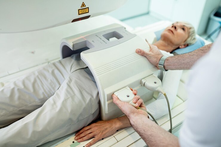
Fluoroscopy-Barium Meal Swallow

A fluoroscopy barium meal swallow is a diagnostic imaging procedure used to assess the esophagus, stomach, and the first part of the small intestine (duodenum). This test helps to visualize the upper gastrointestinal (GI) tract and identify abnormalities such as strictures, tumors, ulcers, or gastroesophageal reflux disease (GERD).
During the procedure, the patient is typically asked to fast for several hours beforehand. The patient then ingests a liquid containing barium sulfate, a contrast material that coats the lining of the digestive tract and enhances visibility on X-ray images. The fluoroscopy machine takes continuous X-ray images as the barium moves through the esophagus and into the stomach and duodenum.
Radiologists observe the movement and behavior of the barium in real-time, allowing them to assess the function and structure of the upper GI tract. Patients may be asked to change positions during the procedure to get different views. After the examination, the patient is usually advised to drink plenty of fluids to help eliminate the barium from their system.
The procedure is generally safe, but some patients may experience mild side effects, such as constipation or abdominal discomfort. It is important for individuals to discuss any pre-existing conditions or concerns with their healthcare provider before undergoing the test.

